Dermoscopy (Dermatoscopy)
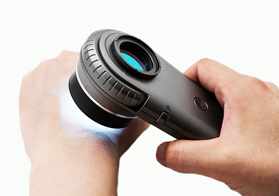
Expert use of dermoscopy (dermatoscopy) has been shown to increase diagnostic accuracy for both melanocytic and non-melanocytic skin malignancies. The evidence for dermoscopy is such that it is now regarded as the standard of care for the evaluation of pigmented skin lesions. Dermoscopy is an advanced examination skill and accredited specialist training is required for effective use.
Our doctors have completed accredited courses in the use of dermoscopy and hold advanced certification in the clinical use of dermoscopy. Dr. Bothma and Dr Coetzer-Botha are members of the International Dermoscopy Society and are enrolled in continuing masterclass education programs.

Mole mapping (Mole photography)
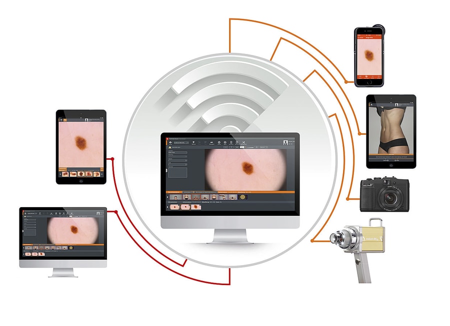
Not all “funny looking” coloured lesions need immediate biopsy or removal. Certain types are monitored at DermaSurg for a specified time frame (usually around 3 – 6 months) using so-called “focused mole mapping” to monitor for changes that might identify those lesions that do require biopsy.
People with extremely high individual risk of developing melanoma are also offered this service in combination with dermoscopy, which remains the gold standard examination to detect early melanoma.

Skin Biopsy
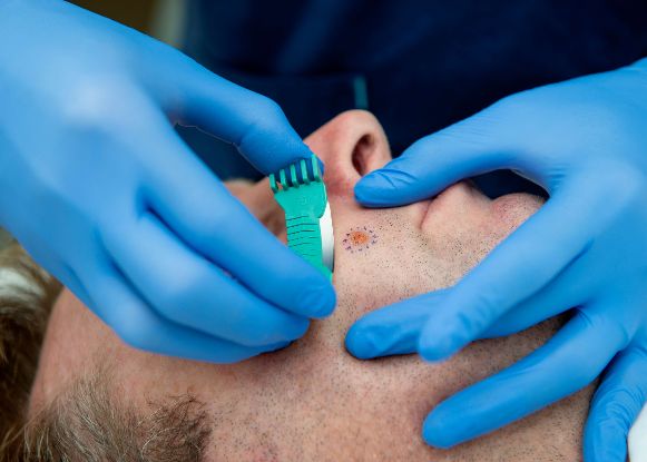
A skin biopsy is one of the most important tools in the investigation and treatment of skin diseases and skin cancers. It involves the removal of a small piece of your skin for examination under a microscope by a specialist dermato-pathologist. The area is first cleaned and then a local anaesthetic is injected under the skin before the procedure is performed. There are several different types of skin biopsy including shave biopsy, punch biopsy, excisional biopsy, and deep shave removal biopsy. Most forms of biopsy require no sutures and heal spontaneously within a few days.
DermaSurg doctors normally perform shave biopsies for most type lesions and deep shave removal biopsies (saucerization) for pigmented (coloured) lesions as they heal fast and leave minimal scarring while providing good samples for pathologists to test. Occasionally an excision biopsy will be indicated and sutures inserted to optimize the healing process.


